Explore the structure and functions of arteries, vital blood vessels that carry oxygenated blood away from the heart. Learn about the layers of arteries – intima, media, and adventitia – and their role in regulating blood flow.
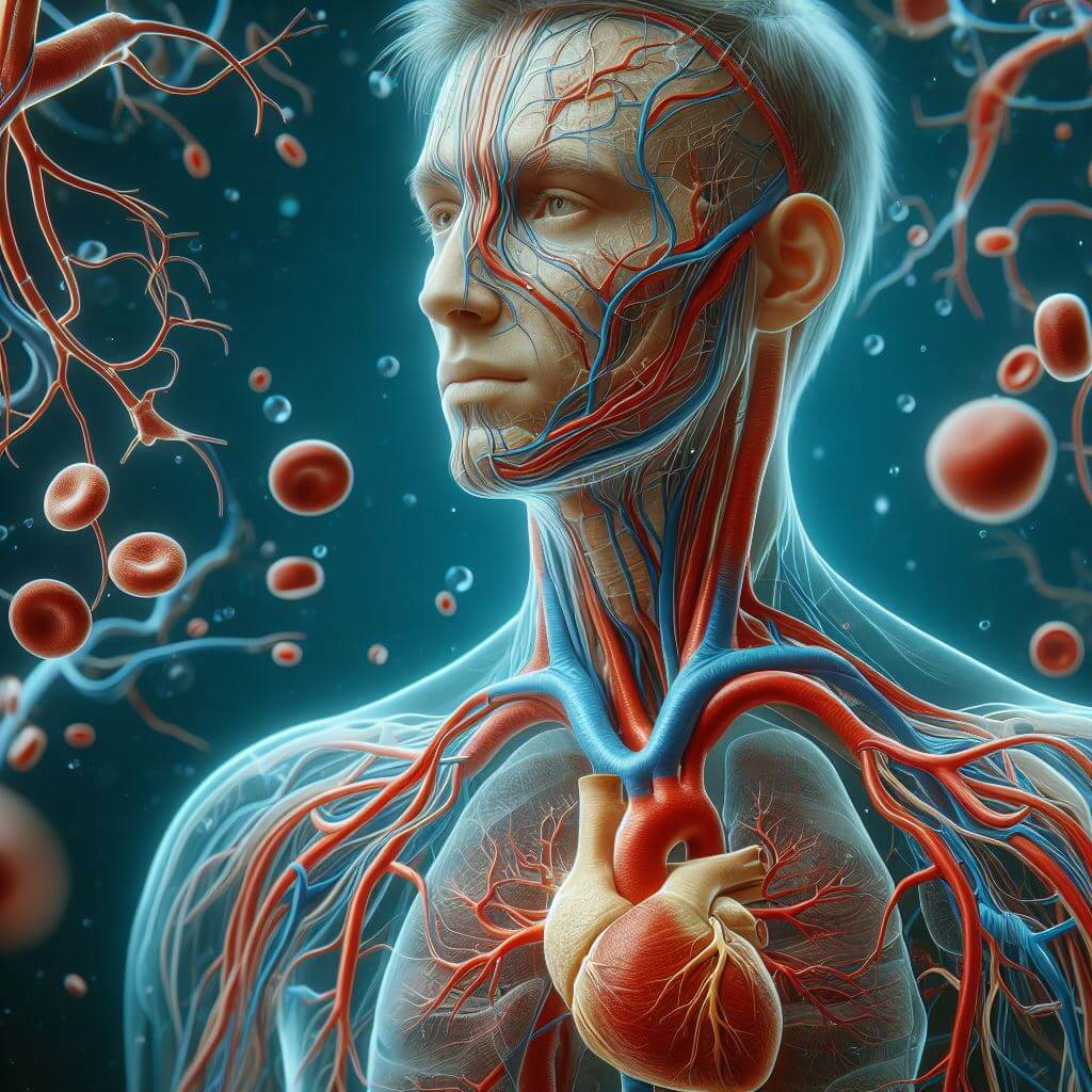
Artery; one of the many blood vessels that carry blood away from the heart to the body cells. The largest artery is the aorta, which begins at the left ventricle of the heart and gives rise to several other arteries as it travels downward through the body. These other arteries soon branch and divide to form smaller arteries, which, in turn, branch and divide to form even smaller arteries. The smallest arteries, arterioles, connect with the capillaries, from which oxygen and food materials in the blood diffuse out to the body cells. At the same time that these materials are leaving the capillaries, carbon dioxide and other waste materials are entering the capillaries. These wastes are then carried back to the heart through a network of veins. These vessels, the arteries, capillaries, and veins, along with the heart, make up the circulatory system.
In addition to distributing blood to the body cells, the arteries help smooth out the flow of blood as it is pumped from the heart. With each contraction of the left ventricle, blood is forced into the aorta with a sudden spurt. To ensure that the body cells receive a rather steady flow of blood, not intermittent spurts, the arteries, especially the aorta and other large arteries, dampen the spurts to a degree by alternately expanding and contracting. As a spurt of blood enters the artery, the artery walls expand, and after the blood flow has ended, they contract to their normal size. By the time the original spurt of blood enters the smaller arteries, it has been evened out to a large degree.
Structure
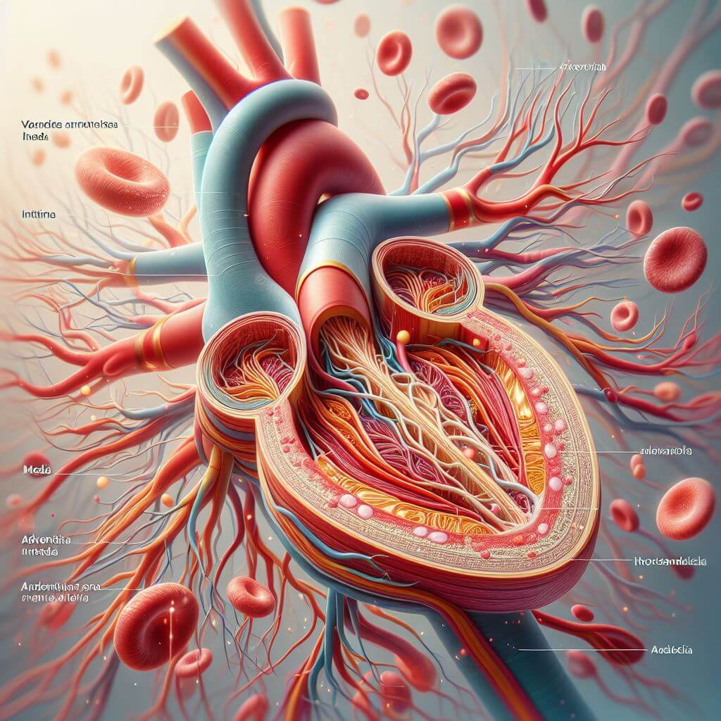
All arteries consist of three basic layers. The innermost layer is called the intima, the middle layer is known as the media, and the outermost layer is the adventitia. Although these three layers are present in all arteries, they vary slightly in thickness and composition among arteries of varying size in the body. Like arteries, veins consist of three layers, but these layers are generally thinner than in arteries. In addition, veins differ from arteries in that the larger ones have flaps of tissue along their innermost surface at various intervals. These flaps, called valves, help keep the blood flowing in the direction toward the heart. Normally the flaps lie flat, but if the blood in a vein starts to back up and flow in the other direction, the flaps of tissue stand up and block the backward flow.
The intima, thinnest layer of the artery wall, consists of three separate sections. The innermost part is made up of endothelial cells, and the middle layer consists of connective tissue, mostly elastic and collagen fibers. In some of the medium-sized arteries this middle section may also have some smooth muscle fibers arranged longitudinally. The fibers are more abundant at the places where the arteries branch. The outermost section of the intima consists of a band of elastic fibers and is called the internal elastic membrane.
The media, usually the thickest layer of the artery wall, consists chiefly of smooth muscle fibers and connective tissue. As the arteries increase in diameter, the layer of muscle tissue becomes thicker. In small arteries the media may consist of only 3 or 4 muscle layers, but in medium-sized arteries there may be as many as 40 layers. The amount of elastic fibers in the connective tissue also varies with the size of the artery. In smaller arteries there are few elastic fibers, and they are scattered among the muscle cells. In medium-sized arteries they are much more numerous and form a circular network that extends throughout the entire media. In the largest arteries the amount of muscle tissue and elastic fibers decreases, and instead of encircling the artery they form bands that wind about in spirals.
The adventitia consists mainly of connective tissue. In small arteries it is a fairly thin layer. In medium-sized arteries it is thick, sometimes as thick as the media, and in the largest arteries it is thin again. The fibers of the adventitia are mostly arranged longitudinally, and the elastic fibers of the innermost section may make up a coarse network, sometimes forming a membrane known as the external elastic membrane. The outermost part of the adventitia consists of connective tissue that is continuous with the connective tissue surrounding the artery.
Diseases
The most common disease of the arteries is atherosclerosis, in which deposits of fatty materials line the inner walls. Other diseases of the arteries include chilblain, frostbite, and Raynaud’s disease.
ARTERIOSCLEROSIS, is a disease that is commonly known as “hardening of the arteries.” The term was applied because, as arteries age and undergo degeneration, calcium is often deposited in their walls, making them hard and brittle. Today, however, the term “arteriosclerosis” has been widely replaced by atherosclerosis, because hardening of the arteries is not always present and is not an important feature of the disease. Atherosclerosis indicates the presence of fatty deposits, called atheromas, on and within the inner layer of the artery wall. These deposits thicken and narrow the arterial channel, thus interfering with the flow of blood. Eventually a clot (thrombus) often forms at the site of the deposit and unless the clot is either dissolved or removed, it may completely obstruct the narrowed artery.
