What is bladder? Information about the general description, structure, functions, diseases of bladder and urination.
The bladder is an essential organ that plays a critical role in the urinary system. It is responsible for storing urine until it is ready to be expelled from the body. Understanding the structure, function, and common disorders of the bladder is essential to maintaining optimal urinary health.
In this post, we will provide a comprehensive overview of the bladder, including its anatomy, function, and role in the urinary system. We will also discuss various disorders and diseases that can affect the bladder, such as urinary tract infections, bladder cancer, and incontinence. Additionally, we will explore the process of urination and the factors that can influence it. By the end of this post, you will have a thorough understanding of the bladder and how to keep it healthy.
Source: wikipedia.org
Bladder; a hollow muscular sac that serves as a reservoir for urine. Urine is formed continuously in the kidneys and is stored in the bladder until it is eliminated. In the average adult, about 1 to 1,5 quarts (0.9 to 1.4 liters) of urine are excreted every day.
General Description
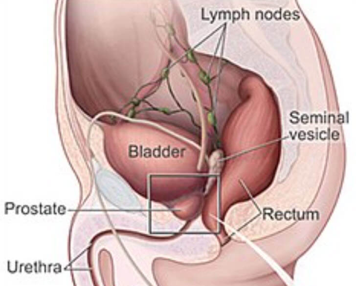
Located in the front section of the pelvis, the bladder is supported and held in place by ligamentous connective tissue. In a man it rests on and is firmly attached to the base of the prostate gland. In a woman it lies just below and in front of the uterus, attached to the cervix (the neck of the uterus) and the front wall of the vagina.
The size and shape of the urinary bladder depends largely on the amount of urine it contains. When empty, it appears as a rounded, deflated balloon, with its inner surfaces flattened against each other. As it fills with urine, the bladder increases in size and becomes pear-shaped. When it is completely filled, the bladder can hold approximately 17 to 18 ounces (500 to 530 ml) of urine.
From each kidney, urine passes into the bladder through a slender 10- to 12-inch (255- to 305-mm) tube called a ureter. Each ureter enters the badder at an oblique angle, allowing the musculature of the bladder wall to act as a sphincter around each opening to prevent the flow of urine from backing up into the ureter.
At the very bottom of the bladder is a thick-walled tube, called the urethra, which conveys the urine to the outside. Immediately beyond the bladder, the urethra passes through a sheet of muscle tissue called the urinogenital diaphragm. At the point where the urethra joins the urinogenital diaphragm, it is surrounded by a thick band of tissue known as the external sphincter. In a man, the urethra is about 8 inches (205 mm) long while in a woman it is only about 1 to 1,5 inches (25 to 38 mm) long. The ‘three openings into the bladder—the two ureters and the urethra —form the three points of an imaginary triangle. The area inside this triangle is sometimes referred to as the vesical trigone.
Structure
The bladder wall consists of three separate layers of tissue. The innermost layer is a mucous membrane that lines the inner surface and is continuous with the membrane that lines the ureters and the urethra. It is a thin membrane, pale pink in color, and it is wrinkled, with many folds.
The next two layers consist of smooth muscle tissue, and the term detrusor muscle is applied to both layers. The innermost layer is a thick circular band of tissue around the bladder, lying transverse to the long axis. At the base of the bladder this muscle tissue forms a dense band around the neck of the urethra. This band is called the internal sphincter. The outer muscle layer is principally longitudinal. When both muscle layers contract, they decrease both the height and the width of the bladder.
Running throughout the layers of the bladder wall are vast networks of lymph and blood vessels. In addition, the bladder is well supplied with both sensory and motor nerve fibers.
Urination
Urination, also called micturition or voiding, is the act of expelling urine from the bladder. As urine accumulates in the bladder, the bladder walls stretch to accommodate this increasing volume. This stretching stimulates sensory nerve fibers in the bladder wall to transmit nerve impulses to the spinal cord. Some of these sensory impulses are then relayed to centers in the brain, where they are interpreted as a desire to urinate.
The act of urination is controlled by both voluntary and involuntary muscles. The voluntary muscle is the external sphincter, which is usually in a relatively contracted state but can be made to relax at will. The involuntary muscle is the detrusor muscle, which is stimulated to contract by impulses from the spinal cord over parasympathetic nerve fibers. These impulses are part of a reflex initiated by the sensory impulses to the spinal cord from the bladder wall.
Diseases
The urinary bladder is susceptible to a variety of diseases and disorders. Its function may also be disturbed by diseases affecting other organs and tissues. For example, diseases of the lower portion of the spinal cord or the nerves leading to or from the bladder may have a marked effect on the frequency of urination.
One of the most common disorders of the bladder itself is cystitis, an inflammation of the bladder usually caused by bacteria. Symptoms of cystitis include increased frequency of urination, sensations of burning during urination, and the passing of cloudy, sometimes blood-tinged, urine. Cystitis is usually treated by the administration of antibiotic drugs such as tetracycline and penicillin.
The bladder is also susceptible to the formation of calculi, or stones, which are hard masses of minerals and salts. Bladder stones may occur singly or in large numbers and sometimes lodge in the opening of the urethra, cutting off the flow of urine. Bladder stones may often be removed with an instrument called a lithotrite, which is inserted into the bladder and manipulated to crush the stones.
Other disorders of the bladder include hernias, fistulas, and malformations of the bladder that are usually present at birth. Cancer of the bladder is relatively rare, occurring mostly in men over the age of 50.
