
Discover the essential features that make microscopes invaluable tools for scientific exploration. Explore magnification, illumination, and more!
The microscope is a tool that allows you to observe objects and elements that are too small to be seen by the naked eye. Its name comes from the Greek “micrós” (diminutive) and “scopéo” (to look), and it uses the principle of the refraction and reflection of the light to generate a controlled increase of the image of the matter.
The microscope was invented in the sixteenth century through an optical system of magnifying lenses, which has been refined to the present day, in which there are so powerful electronic variants that allow us to glimpse the tiniest objects.
This instrument allowed the deep understanding of microscopic life and therefore changed the understanding of life in its entirety, thus becoming an indispensable tool for modern medicine, biology and pharmacology.
Characteristics Of Microscope
1. First microscope
The first microscope is credited to Zacharias Janssen, a Dutch spectacle maker, and his father Hans Janssen in the late 16th century. They developed a simple compound microscope around 1590. This early microscope consisted of two convex lenses within a tube and could magnify objects up to 10 times their actual size. It marked the beginning of a revolutionary era in the world of science and exploration.
2. Evolution
Microscope evolution has been a remarkable journey, leading to the development of increasingly powerful and versatile instruments for scientific research. Here’s a brief overview of the key stages in the evolution of microscopes:
- Simple Microscopes: The early 17th century saw the refinement of simple microscopes with a single lens, thanks to scientists like Robert Hooke and Antonie van Leeuwenhoek. These microscopes provided relatively low magnification but were crucial for observing tiny objects and organisms.
- Compound Microscopes: In the mid-17th century, improvements led to the creation of compound microscopes with multiple lenses. Prominent figures like Galileo Galilei and Johannes Kepler contributed to these advancements. Compound microscopes provided higher magnification and clearer images.
- Brightfield Microscopes: In the 19th century, brightfield microscopes became prevalent. They used a condenser and objective lenses to direct light through a specimen, resulting in enhanced visibility. This era also introduced achromatic lenses, reducing color distortion.
- Phase-Contrast Microscopes: In the early 20th century, the phase-contrast microscope was invented by Fritz Zernike, allowing the visualization of transparent specimens without staining. This was a breakthrough in cell biology.
- Fluorescence Microscopes: In the mid-20th century, the development of fluorescence microscopy enabled the labeling and visualization of specific molecules within cells. This technique revolutionized molecular biology and biomedical research.
- Electron Microscopes: The 1930s saw the birth of electron microscopy, which used electron beams instead of light to achieve much higher magnifications. Transmission electron microscopes (TEM) and scanning electron microscopes (SEM) have since become indispensable tools for nanoscale research.
- Confocal Microscopes: In the 1950s, confocal microscopy emerged, providing three-dimensional imaging capabilities by eliminating out-of-focus light. This technology became crucial in the study of biological tissues and living cells.
- Scanning Probe Microscopes: In the 1980s, the invention of scanning tunneling microscopes (STM) and atomic force microscopes (AFM) allowed scientists to visualize and manipulate individual atoms and molecules at the nanoscale.
- Super-Resolution Microscopes: Recent advances in super-resolution microscopy techniques, such as STED (stimulated emission depletion) and PALM (photoactivated localization microscopy), have broken the diffraction limit, enabling unprecedented clarity at the nanometer level.
- Cryo-Electron Microscopes: Cryo-EM has emerged as a powerful technique for imaging biological macromolecules and cellular structures in their native state, contributing significantly to structural biology and drug discovery.
The evolution of microscopes has played a pivotal role in advancing various scientific fields, from biology and chemistry to materials science and nanotechnology. These advancements continue to drive innovation and exploration at the microscopic and even nanoscopic levels.
3. Parts that compose it
A microscope consists of several key parts and components, each playing a specific role in magnifying and visualizing objects. Here are the primary parts that compose a typical compound light microscope:
Eyepiece (Ocular Lens): The eyepiece is the lens at the top of the microscope that you look through. It typically provides 10x magnification and is where you place your eye to view the specimen.
Objective Lenses: These are the lenses located on the revolving nosepiece just above the specimen. Objective lenses come in various magnifications, such as 4x, 10x, 40x, and 100x (oil immersion). These lenses are responsible for magnifying the specimen.
Revolving Nosepiece: The revolving nosepiece holds the objective lenses and allows you to easily switch between different magnifications by rotating it.
Stage: The stage is a flat platform where you place the specimen slide for observation. It often includes stage clips or a mechanical stage for securely holding the slide in place and moving it in different directions.
Condenser: The condenser is located beneath the stage and helps focus light onto the specimen. It can be adjusted to control the intensity and angle of illumination.
Illumination Source: A light source, typically an adjustable halogen or LED light, provides illumination to the specimen. The light source can be located either above (transmitted light) or below (reflected light) the stage, depending on the type of microscope.
Diaphragm/Iris Diaphragm: The diaphragm is located beneath the condenser and controls the amount of light that reaches the specimen. It can be adjusted to optimize contrast and brightness.
Coarse and Fine Focus Knobs: These knobs are used for focusing the microscope. The coarse focus knob is used for initial rough focusing, while the fine focus knob allows for precise adjustments to bring the specimen into sharp focus.
Base: The base of the microscope provides stability and support for the entire instrument.
Body Tube: The body tube connects the eyepiece to the objective lenses and houses the optical components that transmit the image from the objective to the eyepiece.
Arm: The arm is the curved part of the microscope that connects the body tube to the base. It is used for carrying and holding the microscope.
Stage Height Adjustment: On some microscopes, there may be a mechanism to adjust the height of the stage, allowing for thicker specimens or different observation techniques.
Mechanical Stage Controls: In more advanced microscopes, a mechanical stage may include knobs for precise movement of the specimen slide in both the X and Y directions.
Immersion Oil (for Oil Immersion Objective): If the microscope has a 100x oil immersion objective, immersion oil is used to improve the resolution by reducing the loss of light between the specimen and the lens.
These are the fundamental components of a compound light microscope, which is commonly used in laboratories and educational settings. Electron microscopes and other specialized microscopes have different components and principles of operation.
4. Refraction and Reflection
Refraction and reflection are two fundamental optical phenomena that occur when light interacts with different materials or surfaces. Here’s an explanation of each:
Reflection:
Reflection occurs when light “bounces back” from a surface. When a beam of light strikes a reflective surface, such as a mirror or water, it follows the law of reflection, which states:
- The incident ray (the incoming ray of light) and the reflected ray (the outgoing ray of light) lie in the same plane.
- The angle of incidence (the angle between the incident ray and the perpendicular, or normal, to the surface) is equal to the angle of reflection (the angle between the reflected ray and the normal).
For example, when you look in a mirror, you see your reflection because light from you (the incident ray) strikes the mirror and reflects back towards your eyes (the reflected ray). This phenomenon allows us to see objects and images by bouncing light off surfaces.
Refraction:
Refraction occurs when light changes its direction as it passes from one transparent medium (e.g., air, water, glass) into another with a different optical density. This bending of light is due to a change in its speed as it enters a different medium. The key principles of refraction are:
- When light travels from a less dense medium (lower refractive index) to a denser medium (higher refractive index), it bends toward the normal (an imaginary line perpendicular to the surface at the point of incidence).
- When light travels from a denser medium to a less dense one, it bends away from the normal.
- The degree of bending (the angle of refraction) depends on the difference in refractive indices between the two media and the angle at which the light enters.
Common examples of refraction include the bending of light as it passes through a glass prism, causing the separation of colors in a rainbow, and the way objects appear displaced when viewed through a glass of water.
Refraction also plays a crucial role in the operation of lenses, such as those in eyeglasses and camera lenses, where the bending of light is used to focus images. Additionally, mirages, where distant objects appear displaced or inverted due to atmospheric refraction, are another fascinating example of this phenomenon.
In summary, reflection involves the bouncing back of light from a surface, while refraction involves the bending of light as it passes from one medium into another with a different refractive index. Both phenomena are essential in understanding how light interacts with objects and materials in our daily lives and in various scientific and technological applications.
5. Types of microscopes
There are several types of microscopes, each designed for specific purposes and offering various capabilities. Here are some common types of microscopes:
- Light Microscope (LM):
- Also known as an optical or compound microscope.
- Uses visible light to magnify and observe samples.
- Commonly used in biology and material science for examining biological specimens, cells, tissues, and thin sections of materials.
- Can have multiple objective lenses for different levels of magnification.
- Electron Microscope (EM):
- Uses a focused beam of electrons instead of light to achieve much higher magnification.
- Types of electron microscopes include Transmission Electron Microscopes (TEM) for thin specimens and Scanning Electron Microscopes (SEM) for three-dimensional surface imaging.
- EMs are essential for studying nanoscale structures and materials.
- Scanning Probe Microscope (SPM):
- Includes types like Scanning Tunneling Microscope (STM) and Atomic Force Microscope (AFM).
- Operates by scanning a sharp tip over the surface of a sample and measures various properties at the atomic and molecular levels.
- Used for surface analysis and manipulating individual atoms and molecules.
- Fluorescence Microscope:
- Employs fluorescence to study biological molecules and structures.
- Labels specific molecules with fluorescent dyes or proteins to visualize their location and activity within cells and tissues.
- Widely used in cell biology, genetics, and neuroscience.
- Confocal Microscope:
- Uses a focused laser beam and a pinhole to create sharp, three-dimensional images of fluorescently labeled specimens.
- Provides optical sectioning, eliminating out-of-focus light, which allows for better depth perception.
- Valuable for studying complex biological structures.
- Phase-Contrast Microscope:
- Enhances the contrast of transparent and colorless specimens without staining.
- Relies on differences in refractive index to create contrast in the image.
- Commonly used for live cell imaging.
- Darkfield Microscope:
- Illuminates the specimen with oblique or peripheral light, causing objects to appear bright against a dark background.
- Useful for observing unstained or weakly stained specimens and for detecting very small objects.
- Polarizing Microscope:
- Analyzes the optical properties of minerals and materials by examining their response to polarized light.
- Widely used in geology and material science to identify and characterize crystalline structures.
- Inverted Microscope:
- Designed with the light source and condenser above the specimen stage.
- Ideal for observing living cells and tissues in culture dishes or flasks.
- Digital Microscope:
- Integrates a digital camera for capturing images and videos of specimens.
- Allows for easy sharing and analysis of images on a computer screen.
- Commonly used in educational settings and for documentation.
These are some of the main types of microscopes, each tailored to specific research or observation needs in fields ranging from biology and chemistry to materials science and nanotechnology.
6. Optics and lenses
Optics and lenses play a fundamental role in the science of optics and various optical devices, including microscopes, telescopes, cameras, and eyeglasses. Here’s an overview of optics and lenses:
Optics: Optics is the branch of physics that studies the behavior of light, including its properties, interactions, and how it travels. It encompasses various phenomena related to the behavior of light, such as reflection, refraction, dispersion, diffraction, polarization, and more. Optics is essential in understanding how light interacts with matter and how it can be manipulated to create various optical devices and technologies.
Lenses: Lenses are optical components made of transparent materials, typically glass or plastic, that have curved surfaces. Lenses are designed to refract (bend) and focus light, either converging it to a point or spreading it out. There are two primary types of lenses:
- Convex Lenses:
- Convex lenses are thicker in the center and thinner at the edges.
- They converge parallel rays of light and are often called converging lenses.
- Convex lenses can be used to form real or virtual images, depending on the position of the object relative to the lens.
- Concave Lenses:
- Concave lenses are thinner in the center and thicker at the edges.
- They diverge parallel rays of light and are known as diverging lenses.
- Concave lenses always form virtual, upright, and diminished images.
Lenses are classified based on their shape and purpose:
- Double-Convex (Bi-Convex) Lens: Both surfaces are outwardly curved.
- Double-Concave (Bi-Concave) Lens: Both surfaces are inwardly curved.
- Plano-Convex Lens: One surface is flat (plano), and the other is outwardly curved.
- Plano-Concave Lens: One surface is flat (plano), and the other is inwardly curved.
- Meniscus Lens: One surface is convex, and the other is concave.
Lenses have various applications:
- Eyeglasses: Convex and concave lenses are used to correct vision problems like nearsightedness and farsightedness.
- Microscopes: Compound microscopes use multiple lenses to magnify small objects for detailed examination.
- Telescopes: Refracting telescopes and reflecting telescopes utilize lenses and mirrors to gather and focus light for astronomical observation.
- Cameras: Camera lenses focus light onto a photosensitive surface (film or sensor) to create images.
- Projectors: Lenses are used to project images or videos onto screens or surfaces.
- Optical Instruments: Lenses are crucial in various scientific instruments, including spectrometers, spectrophotometers, and more.
- Magnifying Glasses: Convex lenses are used in magnifying glasses to enlarge small objects for better viewing.
- Lasers: Lenses are used to shape and focus laser beams for various applications, from surgery to data transmission.
Understanding optics and the behavior of light through lenses is essential for designing and optimizing optical systems, whether for scientific research, healthcare, photography, or telecommunications. The study of optics and the use of lenses have significantly advanced technology and our understanding of the physical world.
7. Preparation of the object
To use the microscope, we must prepare the object to be observed, mounting it on a transparent glass sheet called a slide. Then it is covered with another thinner and smaller lamella called coverslip and, if necessary, stains or dyes are added to identify what is sought.
8. Virtual microscopes
This is called a novel technique for analyzing data areas of a cytological or histological sample (cells and tissues), using a computer system that reproduces the information captured in a simulated environment.
This technique is in development in current times, since it would allow the full integration of computer systems to scientific research or its transmission over long distances and in real time, taking advantage of new Internet-inspired technologies.
This is also a step towards the automation of certain processes of scientific analysis that could dispense with the constant intervention of man, such as histological examinations or blood samples.
9. Importance for medicine
The birth of microscopy and the use of this tool in the various life sciences prompted a scientific revolution that was already insinuated in theoretical terms.
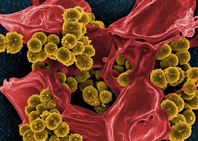
The verification of smaller life forms of the visible abolished magical or religious theories about the disease altogether, and opened a scientific window for the verifiable study of the different infectious agents (viruses, bacteria, protozoa) and chemistry own body (blood tests).
10. Care
A microscope should be used with care to avoid damaging its delicate functioning system, such as carrying it with both hands, since a fall is fatal; do not move it while it is used; do not touch the lenses or the eyepiece, as they get dirty easily; or avoid for the same reasons that the end of the lens touches the observed substance when approaching it to focus.
